
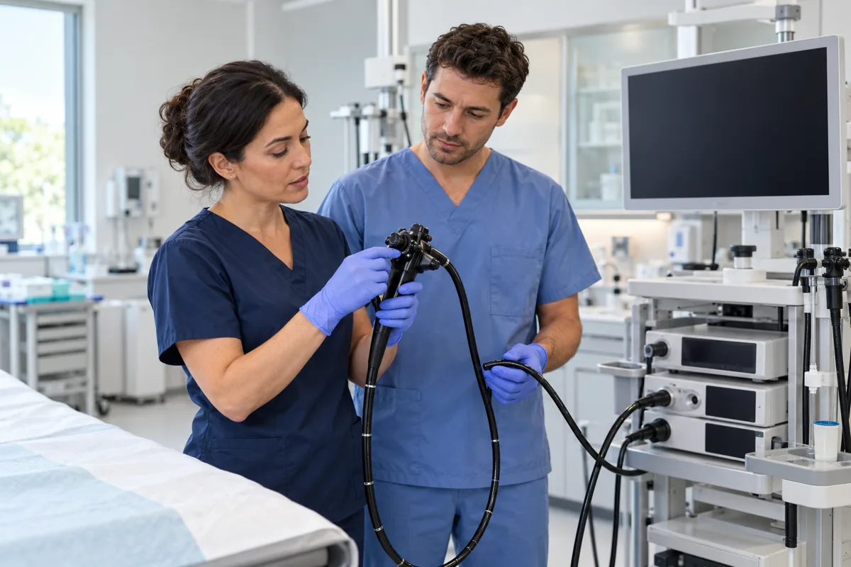
What HD and NBI mean
High-definition, or HD, imaging improves the detail displayed during endoscopy. Narrow band imaging, or NBI, uses selected narrow bands of light to accentuate superficial blood vessels and mucosal surface patterns. The endoscopist can switch between white light and enhanced views during the same examination. These technologies are components of an endoscopy system, rather than a CT scan or an independent treatment. They do not rely on X-rays or radioactive substances.
When enhanced views can help
Enhanced imaging can help identify subtle abnormalities, assess the borders of a lesion, target biopsies and characterise selected polyps. Its usefulness depends on the organ, indication, operator experience and relevant guideline. Selected oesophageal, gastric and colorectal surveillance examinations may benefit. Availability does not mean that every colour change needs treatment or that every examination requires extra interventions. The purpose is to support better decisions in appropriate settings.
Why images do not settle every diagnosis
Neither HD nor NBI guarantees detection of every lesion or confirms cancer simply from its appearance. Good bowel preparation, clear mucosal views, careful inspection, training and tissue examination when indicated remain essential. A report may describe a suspicious pattern requiring a biopsy, removal or a specific surveillance plan. The words “advanced imaging” do not make a finding final. Optical diagnosis strategies for small polyps require specific quality and training conditions and should not be assumed to apply universally.
Preparation, risks and the patient experience
Preparation follows the underlying procedure: fasting and medication instructions for upper endoscopy, bowel cleansing for colonoscopy, and an escort and driving restrictions when sedation is used. Switching on NBI does not itself require a universal additional preparation programme. Patients usually do not feel the light change. Risks remain those of endoscopy, sedation and any biopsy or polyp removal, including bleeding and rare perforation. Ask which findings were seen, whether tissue results are pending and how follow-up will be arranged. Severe pain, fever, substantial bleeding or breathing problems afterwards need urgent contact with the team.
Preparing for a clinical appointment
Once any urgent symptoms have been addressed, prepare a clear description of the problem rather than relying on a diagnosis you suspect. Record when symptoms started, how often they occur, any relationship to meals, sleep or medicines, and their effect on daily activities. Bring previous results and a list of medicines, supplements and allergies. Mention chronic illnesses, operations and relevant family history. This information helps guide the next step and may avoid unnecessary repeat investigations. Never delay seeking care while collecting paperwork or completing a diary.
Understanding investigations and treatment
Ask what each test is intended to establish: a cause, a complication or a response to treatment. How would its result change the plan? Different patients need different investigations. A normal result does not make symptoms unreal, and an abnormal result needs interpretation in context. If medication is prescribed, clarify its name, timing, duration, important adverse effects and instructions for a missed dose. Do not borrow another person’s prescription, start antibiotics or stop long-term treatment independently.
Making follow-up practical
Agree when review should occur, how results will be communicated and who will answer questions. Ask what to do if symptoms persist or change, and which developments warrant earlier contact. Tell the team if cost, transport, laboratory access or work makes the plan difficult to follow so that suitable alternatives can be considered. The aim is a useful explanation and a workable care plan, rather than the largest possible number of tests.
Write down two or three priority questions and ask for unfamiliar terms to be explained. A trusted companion can help you remember instructions if you wish, while respecting your privacy. Avoid highly restrictive diets or supplements advertised as cures without reliable evidence. Discuss major dietary changes with an appropriate professional. Symptom improvement matters, but it does not replace tests or monitoring requested for a particular reason. Keep an accessible summary of the plan and results to support continuity if you attend another clinic.
Scientific sources
ESGE — Advanced imaging guideline update 2019
ESGE — Advanced imaging in the upper GI tract
Medical notice: This information is for health education and does not replace medical consultation or individual assessment. Seek urgent care for warning signs.